Search

12 Aralık 2015 Cumartesi

Histiocytic Nodules


  • Nodular aggregates of histiocytes resembling Langerhans'/interdigitating cells can occur in the  endometrium. The examples described by Kim et al. were an incidental microscopic finding in curettage specimens in women of reproductive age. The nodules were solitary and 0.3-1.5 cm in maximum.
  • The histocytes are round to polygonal with distinct cytoplasmic borders and moderate amounts of pale amphophilic or eosinophilic granular cytoplasm. Small cytoplasmic vacuoles, but not cytoplasmic lipid or pigment, may be present. The avoid to reniform nuclei have occasional grooves, fine chromatin, and inconspicuous nucleoli. Mitoses were frequent in one case.
  • The cells are immunoreactive for CD68 and lysozym but negative for S100 and cytokeratin.
  • Uterine involment by Langerhans' cell histocytosis can be excluded by the absence of eosinophils as well as negative staining for S100. The distinctive apperance of the histiocytes, their nodular arrangement, and the absence of lipid and/or pigment xanthogranulomatous endometritis.

MONODERMAL TERATOMAS


Struma Ovarii


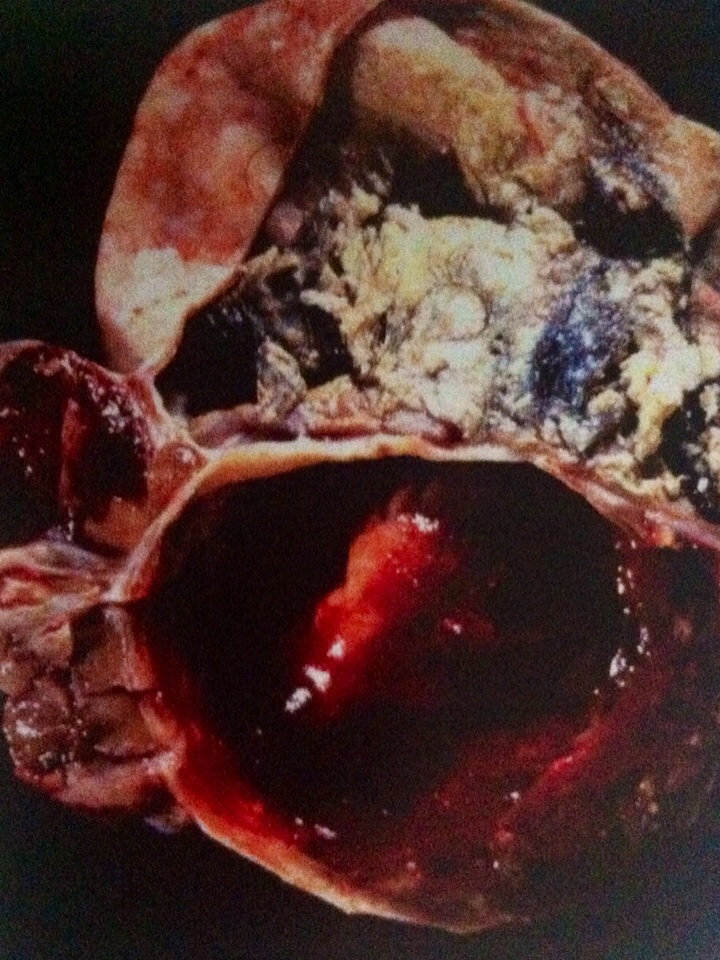
  • The term 'struma' is reserved for cases in which thyroid tissue is the predominant or sole component or forms a grossly recorgnizable tumor in a teratoma.
  • The peak frequency is in the fifth decade, but occasional cases occur much earlier or later. When clinically significant (many are incidental findingsin a resected dermoid), the presentation is similar to that of any ovarian mass. In rare cases, however, there is clinical evidence of hyperthroidism.
  • Ascites is present in as many as a third of cases, and occasionally is accompanied by Meigs' syndrome. The association of a pelvic mass, ascites, and an elevated serum level of CA-125 can mimic ovarian cancer.

10 Aralık 2015 Perşembe

PAPILLARY PROLIFERATION


  • Lehman and Hart have describe localized papillary endometrial proliferations that could lead to a misdiagnosis of a villoglandular endometrioid adenocarcinoma,especially in a fragmented biopsy or curettage specimen.
  • The lesions typically occur in postmenopausal women who present with bleeding. Two-thirds of the lesions involved polyps, but in some cases the lesion occured in the absence of, or at a distance from, a polyp.
  • The proliferations are characterized by papillae with fibrovascular cores and variable degrees of branching. The papillae are covered by epithelial cells with bland to mildly atypical nuclei, with occasional mitotic figures in some cases.
  • One or more metaplastic epithelial changes are often present, including (in descending order of frequency in the cited study) mucinous, eosinophilic cell, ciliated cell, squamous, and hobnail cell metaplasia.
  • The patients had uneventful outcomes, but only three who were not treated by hysterectomy had appreciable follow-up.
  • Awareness that the lesion tend to occur in polyps, the typically bland cytologic features, and the usual presence of metaplastic epithelia facilitate the diagnosis.

ENDOMETRİAL POLYPS


  • Endometrial polyps are typically encountered in the reproductive and postmenopausal age groups;65% of patients are over the age of 40. Polyps are present in as many as 25% of endometrial biopsy specimens performed for abnormal uterina bleeding, although they are often an incidental finding. Sbouth 10%of polyps prolapse into the endocervix, often mimicking an endocervical polyp.
  • Polyps are the most common endometrial lesion associated with tamoxifen therapy, being present in as many as one-third of these patients. Tamoxifen-related polyps tend to be larger, more commonly multiple, and are more likely to recur.
  • Polyps most often arise in the fundus. In a study of 1100 polyps, Peterson and Novak found them to range from 0.3 to 12 cm (mean, 2.3 cm); 20% werw multiple. Polyps have a broad to narrow base, a usually smooth external surface, and an often cystic and/or fibrotic sectioned surface. Focal hemorrhage may be seen, particularly at their tips, due to torsion with subsequent infaretion.
Microscopic features
  • The glandular and stromal morphology is highly variable and may differ from or reflect the appearance elsewhere in the endometrium.
  • The glands are often inactiveand cystic, particularly after the menopause, but functional (proliferative or secretory), metaplastic, hyperplastic, or even carcinomatous glands may be present. The glands are often arranged parallel to the surface epithelium. We allow some gland crowding in polyps, including cellular stratification and mitotic activity, withouth rendering a diagnosis of hyperplasia.
  • A mixture of endometrial and endocervical-type glands may be ancountered in polyps arising near the internal os.
  • Glandular features more commonly present in tamoxifen-related than in tamoxifen-unrelated polyps includeglands polarized along the long axis of the polyps, focal stromal hypercellularity, staghorn-shaped glands, small glands, and metaplastic glands.
  • Polyps may contain a distinctive proliferation of fibrous-cored papillae and metaplastic epithelium, as described by Lehman and Hart (see below).
  • Hyperplasia and adenocarcinoma have been found in as many as 11-30% and 0.5-3% of polyps, respectively, in the general population. Carcinoma has been found in 3.0- 10.7% of polyps in tamoxifen-treated women.
  • İn women with complex hyperplasia within polyps, da Costa and Mittal found hyperplasia or adenocarcinoma of nonpolypoid endometrium in the hysterectomy specimen in 72% and 31% of cases, respectvely. Some of the carcinomas were myoinvasive.
  • Carcinomas found in polyps (tamoxifen related and tamoxifen unrelated) may be confined to the polyp or be part of a multifocal endometrial neoplasia. They are usually endometrioid or serıous, or rarely other cell types. The serous carcinomas, in particular, are often microscopic and /or noninvasive and can be overlooked. Tmoxifen-related polyps may also harbor metastatic lobular breast carcinoma.


  • The stroma of endometrial polyps may be fibrotic and sparsely cellular, especially in tamoxifen-related polyps. İn other cases, it resembles normal or hypercellular endometrial stroma; in these polyps, mitotic figures in the stromal cells may be present.
  • Blood vessels, which are often thick-walled and/or hyalinized, are more numerous in polyps than in the normal endometrium, and are sometimes a helpful clue to the diagnosis.
  • Atypical stromal cells, which may be multinucleated, are a rare focal, multifocal, or diffuse finding within a polyp. Their nuclei can be moderately to severely atypical and hyperchromatic, often with a smudged appearnce. Mitotic figures are usually absent.
  • Other uncommon stromal findings include foci of smooth muscle and decidua, the latter usually reflecting exogenous progestin use latter usually reflecting exogenous progestin use or pregnancy.
  • Vascular thrombosis and hemorrhagic necrosis can occur within the polyp, and hemorrhagic necrosis can occur within the polyp, and may result in reactive atypia of the epithelium or stroma of the polyp.

9 Aralık 2015 Çarşamba

CELLULAR ANGİOFİBROMA


  • This is an uncammon distinctive soft tissue tumor that in females most commonly arises in the vulva rare tumors have arisen in the vagina, parineum, inguinal region, or urethra.
  • The patents are of peproductive or postmenopausal age (mean,46 years) and present with a superficial mass. The tumors are successfully treated by local excision; one recurred locally.
  • In the largest series, the tumors measured 0.6-12 (median 2.8) cm and were usually well circumscribed, with a solid, white, tan, or grey sectioned surface.
  • Microscopic examnation reveals a cellular proliferation of spindle cells arranged in short interesting fascicles, numerous small to medium-sized, thick-walled, often hyalinized, blood vessels, and short wispy collagen bundles. The spindle cells have scanty eosinophilic cytoplasm and bland nuclei. Mitotic figures are usually uncommon.
  • Fat is present in about 25& of cases, but usually accounts for less than 5% of the tumor. Hypocellular areas related to areas of edema, myxoid change, or hyalinization may be present.
  • Unusual to rare findings include an infiltrative border, absence of thick-walled vessels, dilated hemangiopericytomatous vessels, vague nuclear palisading, mild cytologic atypia, multinucleated cells (like those in symplastic leiomyomas), frequent mitotic figures (up to 11 MFs/10 HPFs), and stromal lymphoid aggregates.
  • The spindle cells are immunoreactive for vimentin, CD34, smooth muscle actin, and desmin in approximately 100%, 50%, 20&, and 10% of caces, respectively, Nuclear staining for ER, PR, or both is present in about half the caces. Staining for keratin, EMA, and S100 protein is negative.


14 Kasım 2014 Cuma

Ksilen


 Ksilen, benzene iki adet metil grubu bağlanmasıyla olusan bileşiktir. Disubstitue benzen olduğundan adlandırılması da benzerdir. Orto ksilen, meta ksilen ve para ksilen olmak uzere üç çeşittir. Toluenle akraba sayılır. Uçucu organik bileşiklerdendir. Zehirli, kanserojen bir bileşiktir. formülü C8H10 dur ve molekül ağırlığı 106.16g/mol. Oda koşullarında renksiz bir sıvıdır.

- O-Ksilen nin yoğunluğu 0.88g/mL ve erime noktası -25C , kaynama noktası 144C dir.
- M-Ksilen nin yoğunluğu 0.86 g/mL ve erime noktası -48C , kaynama noktası 139C dir.
- P-Ksilen nin yoğunluğu 0.86g/mL ve erime noktası -13C , kaynama noktası 138C dir.

Ksilen çok çabuk alev alır, solunması ve deri ile temasından kaçınılmalıdır tahriş edicidir. gözle temasından kaçınılmalıdır. Temas halinde bol su ile yıkanmalıdır.

27 Eylül 2014 Cumartesi

Adli Tıp Patolojisi

      Ölüm Kavramı
    19 .yy'a varıncaya kadar ölüm ile ilgili olarak bilimsel açıdan yeterli bir tanımlama yapılmamıştır. İlk kez Fransız bilim insanı Emanuela Fodere ''somatik ölüm'' (vücut ölümü) tanımını ortaya atmıştır. Somatik ölüm temel vücut fonksiyonları olarak kabul edilen merkezi sinir sistemi,solunum ve dolaşım fonksiyonlarının irreversible kaybıdır. Bu fonksiyonlardan birinin kaybı, otomatikmen kısa bir süre sonra diğerlerinin de kaybını doğuracaktır. Somatik ölüm tanımı, hukuken geçerli olan ölüm tanımıdır; nasıl ki kişinin hukuki varlığı canlı doğması ile başlıyorsa, sona ermeside somatik ölüm tanımı ile olmaktadır. Somatik ölümle birlikte özellikle beyin sapındaki solunum ve dolaşım merkezinin devre dışı kalması sonucu süreç kaçınılmaz olarak tüm organ ve dokuların canlılık durumunu yitirmesi ile sonuçlanacaktır. Buna hücresel ölüm (biyolojik ölüm) denmektedir. Bu ölüm şeklinin, hukuki açıdan bir önemi bulunmaz. Yani, ölen kişi ile ilgili bir takım yasal işlemlerin yapılabilmesi için, diğer organ ve dokuların canlılık durumunu yitirmeleri beklenmez.

   Günümüzde belli durumlar için, somatik ölümle ilişkili ''beyin ölümü'' tanımı kullanılmaktadır. Beyin ölümü kavramı, 20.yy da tıptaki ilerlemelere bağlı olarak ortaya çıkmıştır. Önce, solunum ve dolaşımın yapay aygıtlarla desteklenmesi ile yaşam süresinin uzatılması, sonra kab grubu ve subgruplarının bulunması, antibiyotiklerin bulunması, bağışıklık sistemi ile ilgili buluşlar ile başka bir vericiden doku ve organ transplantasyonu olanağı doğmuştur.